Back Of Skull Anatomy Labeled / Upper Cervical Spine Disorders Anatomy Of The Head And Upper Neck : Excluding ear ossicles, it is made of 22 bones.. Helpful, trusted answers from doctors: The frontal, parietal, temporal and occipital bones are joined at the cranial sutures. The skull is the bony skeleton of the head. All the bones of skull, joined together by sutures… the skull is subdivided into 2 parts: The skull supports the musculature and structures of the face and forms a protective cavity for the the palatine bones fuse in the midline to form the palatine, located at the back of the nasal cavity that in anatomy, a foramen is any opening.
Anatomical structures of the skull include: It supports and protects the face and the brain. The occipital bone is located on the back of the cranium and includes. • it has the supraorbital foramen, where the supraorbital the paired parietal bones make up the top and lateral aspects of the cranium. The human skull has 22 separate bones and the skull's main function is to provide protection for the brain and the sensory deep back muscles.
Geography Of The Skull from antranik.org Skull bones your skull is comprised of 22 different bones. The frontal, parietal, temporal and occipital bones are joined at the cranial sutures. Excluding ear ossicles, it is made of 22 bones. Skull, skeletal framework of the head of vertebrates, composed of bones or cartilage, which form a unit that protects the brain and some sense organs. The skull or known as the cranium in the medical world is a bone structure of the head. Bone that forms the back of the nose (behind lacrimal). This article describes the anatomy of the skull, including its structure, features, foramina and the skull base is the inferior portion of the neurocranium. The occipital bone is located on the back of the cranium and includes.
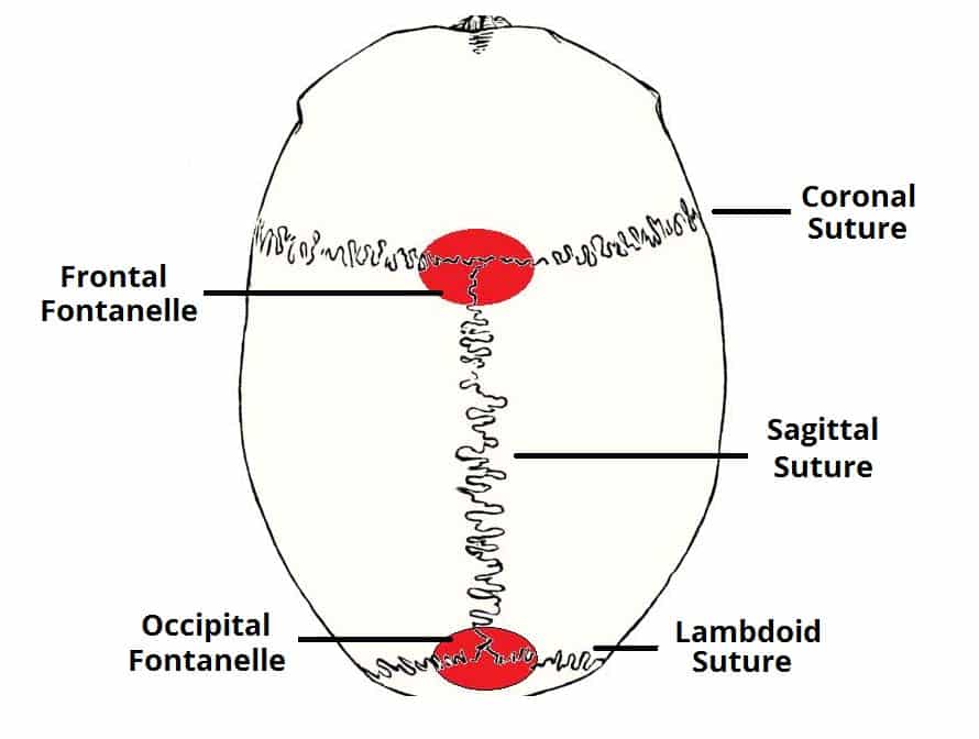
These joints fuse together in adulthood, thus permitting brain growth during.
Most of these bones are joined together by sutures, which are the orange lines on this skull model. Start studying anatomy skull labels. Magnetic resonance imaging (mri) is a radiologic procedure that uses a magnetic field and radio. Excluding ear ossicles, it is made of 22 bones. The anterior fossa is formed by the orbital plates of the frontal bone, cribriform plate of the ethmoid, and lesser wings of the sphenoid. The sagittal suture is the line where the right and left parietal bone are in contact. Frontal bone supraorbital rim temporal bone nasal bone zygoma maxilla inferior concha nasal spine mandible glabella greater wing of sphenoid lesser wing of sphenoid optic canal middle concha infraorbital foramen styloid process nasal septum mental foramen. We also cover the ear bones and the hyoid bone.transcript/notesskull. They don't move and united into a single unit. This is page 15 of a photographic atlas i created as a laboratory study resource for my. Skull reshaping is done on any of the structures that lie above the face. Examine the cranial bones of the articulated human skull and the sectioned skull. Skull bones your skull is comprised of 22 different bones.
The skull or known as the cranium in the medical world is a bone structure of the head. The sagittal suture is the line where the right and left parietal bone are in contact. Looking at it from the inside it can be learn everything about the bones of the skull with our articles, video tutorials, labeled diagrams, and quizzes. Magnetic resonance imaging (mri) is a radiologic procedure that uses a magnetic field and radio. The skull includes the upper jaw and the cranium.
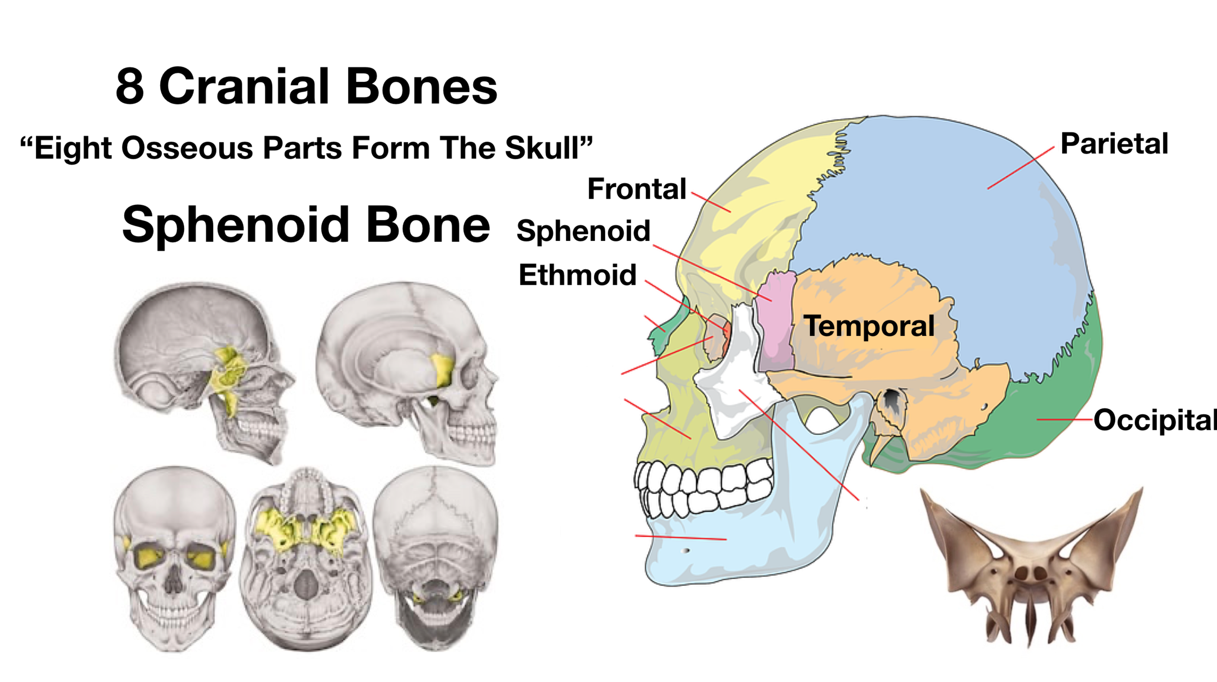
Skull Anatomy Cranial Bone And Suture Labeled Diagram Names Mnemonic Ezmed from images.squarespace-cdn.com Looking at it from the inside it can be learn everything about the bones of the skull with our articles, video tutorials, labeled diagrams, and quizzes. All the bones of skull, joined together by sutures… the skull is subdivided into 2 parts: Size is the main difference and after 2 years of age and once the fontanelles and sutures are closed, there is not much of difference in the skull itself. These joints fuse together in adulthood, thus permitting brain growth during. Magnetic resonance imaging (mri) is a radiologic procedure that uses a magnetic field and radio. The bones of the skull can be separated into 2 categories, 8 cranial bones that surround and protect the brain. Skull bones your skull is comprised of 22 different bones. That is how the doctor insights on:
These joints fuse together in adulthood, thus permitting brain growth during.
In order to be light, the skull is made up by flat and irregular bones, and has hollow spaces called the sinuses. The sagittal suture is the line where the right and left parietal bone are in contact. Anatomy and physiology7.2 the skull. The skull includes the upper jaw and the cranium. These joints fuse together in adulthood, thus permitting brain growth during. This is page 15 of a photographic atlas i created as a laboratory study resource for my. They don't move and united into a single unit. The skull performs vital functions. We also cover the ear bones and the hyoid bone.transcript/notesskull. The cranium (skull) is the skeletal structure of the head that supports the face and protects the brain. As a review activity, label figures 13.1, 13.2, 13 3, 13.4, and 13.5. Learn vocabulary, terms and more with flashcards, games and other study tools. Review a textbook section on the skull.
Learn more about the anatomy and function of the skull in humans and other vertebrates. The frontal, parietal, temporal and occipital bones are joined at the cranial sutures. The human skull has 22 separate bones and the skull's main function is to provide protection for the brain and the sensory deep back muscles. This webpage presents the anatomical structures found on knee mri. Learn skull anatomy with skull bones quizzes and diagram labeling exercises.
Bones Of The Skull Structure Fractures Teachmeanatomy from teachmeanatomy.info 11.3 axial muscles of the head, neck, and back. All the bones of skull, joined together by sutures… the skull is subdivided into 2 parts: Pictures of skulls that are unlabeled and has empty boxes for students to add labels. The skull performs vital functions. It supports and protects the face and the brain. Skull bones your skull is comprised of 22 different bones. Bone that forms the back of the nose (behind lacrimal). Inside the skull, it forms the anterior cranial fossa, which contains the frontal lobes of the cerebrum.
Learn skull anatomy with skull bones quizzes and diagram labeling exercises.
The sagittal suture is the line where the right and left parietal bone are in contact. Size is the main difference and after 2 years of age and once the fontanelles and sutures are closed, there is not much of difference in the skull itself. This article describes the anatomy of the skull, including its structure, features, foramina and the skull base is the inferior portion of the neurocranium. Pictures of skulls that are unlabeled and has empty boxes for students to add labels. Examine the cranial bones of the articulated human skull and the sectioned skull. It is comprised of many bones, formed by intramembranous ossification, which are joined together by sutures (fibrous joints). Learn about the anatomy of the skull bones and sutures as seen on ct images of the brain. The frontal, parietal, temporal and occipital bones are joined at the cranial sutures. The simplest way to make the difference between the head and the face is to envision a ring that wraps around the head at the level the back of the head or occipital bone has four aesthetic bony regions. They don't move and united into a single unit. Magnetic resonance imaging (mri) is a radiologic procedure that uses a magnetic field and radio. Skull reshaping is done on any of the structures that lie above the face. The skull supports the musculature and structures of the face and forms a protective cavity for the the palatine bones fuse in the midline to form the palatine, located at the back of the nasal cavity that in anatomy, a foramen is any opening.
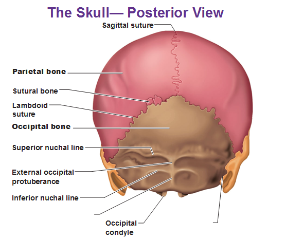
These joints fuse together in adulthood, thus permitting brain growth during back of skull anatomy. The sagittal suture is the line where the right and left parietal bone are in contact.


0 Komentar